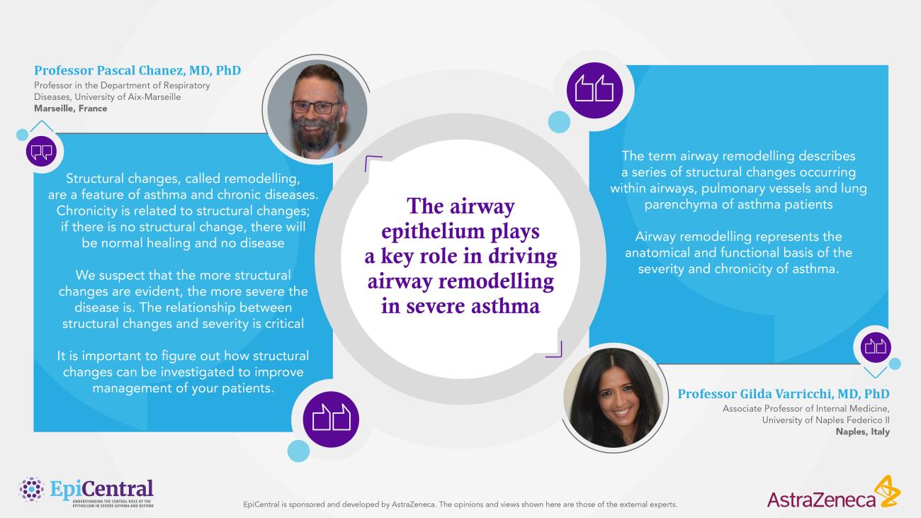
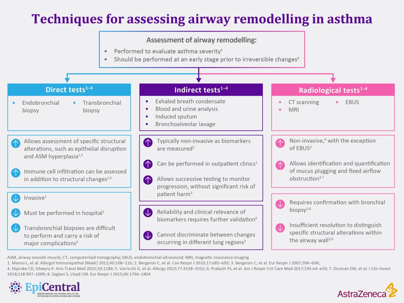
The airway epithelium plays a key role in driving airway remodelling in severe asthma

As an initiator of airway remodelling, the airway epithelium plays a pivotal role in driving structural changes that contribute to the onset and progression of severe asthma.1–4

As an initiator of airway remodelling, the airway epithelium plays a pivotal role in driving structural changes that contribute to the onset and progression of severe asthma1–4

  • Airway remodelling describes heterogeneous structural changes occurring within the small and large airways of patients with asthma1,5,6
  • As the first line of defence against the external environment, the airway epithelium initiates airway remodelling in response to damage by releasing mediators including epithelial cytokines TSLP, IL-33 and IL-251,6,7
  • Epithelial cytokines play diverse, yet often overlapping, roles in airway remodelling through effects on structural and immune cells8
  • Airway remodelling can occur early in life before the onset of symptoms;1,9,10 over time, accumulative remodelling results in structural changes that may impact clinical outcomes in severe asthma1,10
  • Unchecked airway remodelling ultimately results in fixed airflow limitation and contributes to lung function decline, severity, chronicity and fatality1,4,9,11,12

Early identification of airway remodelling, before the onset of significant irreversible structural changes, may aid in clinical decision making and the achievement of disease remission as a goal in severe asthma care9,13–15

1. Hough KP, et al. Front Med (Lausanne) 2020;7:191. 2. Yang Y, et al. Clin Respir J 2021;15:1027–1045. 3. Beckett PA, Howarth PH. Thorax 2003;58:163–174. 4. James AL, Wenzel S. Eur Respir J 2007;30:134–155. 5. Hsieh A, et al. Front Physiol 2023;14:1113100. 6. Varricchi G, et al. Allergy 2022;77:3535–3552. 7. Samitas K, et al. Allergy 2018;73:993–1002. 8. Gauvreau GM, et al. Allergy 2023;78:402–417. 9. Thomas D, et al. Eur Respir J 2022;2102583. 10. Fehrenbach H, et al. Cell Tissue Res 2017;367:551–569. 11. Brightling CE, et al. Clin Exp Allergy 2012;42:638–649. 12. Krings JG, et al. J Allergy Clin Immunol 2021;148:752–762. 13. Zhang J, Dong L. J Thorac Dis 2020;12:6090–6101. 14. Gras D, et al. Med Sci (Paris) 2011;27:959–965. 15. Gupta S, et al. Chest 2009;136:1521–1528. 

Image
Expert Quotes - Professor Chanez and Varricchi

Insights from our EpiCollaborators

What is the clinical significance of airway remodelling in asthma?

Clinically, airway remodelling has implications for the severity of asthma, lung function decline, the risk of future exacerbations and asthma fatality.4,12 As a longitudinal process, airway remodelling is thought to also play a part in the chronicity of asthma.1,10,11

Unchecked airway remodelling results in fixed airflow limitation.1,11 Early identification of airway remodelling, before the onset of significant irreversible structural changes, may therefore aid in clinical decision making and improve disease outcomes in patients with asthma.13–15

The physiology of airway remodelling

Tissue remodelling is a normal physiological response necessary for the resolution of transient cell and tissue damage.10 In patients with asthma this response is aberrant due to contributing factors such as chronic airway inflammation and epithelial abnormalities.16,17 Improper repair in the face of recurrent damage can therefore lead to pathological airway remodelling.1,10,18

Airway remodelling describes structural changes occurring within the small and larger airways of patients with asthma.1,5,6 Physiological changes include epithelial disruption, goblet cell hyperplasia and submucosal gland enlargement, thickening and fibrosis of the subepithelial matrix, angiogenesis and increased airway smooth muscle (ASM) mass.5,6

Airway remodelling is a continuous process, often occurring early in life and persisting through to adulthood.1,3,19,20 Early structural changes are thought to predispose individuals to developing asthma, possibly by affecting structural development of the lungs.10 Cumulative remodelling throughout life can then manifest in the significant structural changes seen in severe asthma.21 Like inflammation, airway remodelling in asthma is heterogeneous5,11,13 and may contribute to the variability seen in asthma phenotypes and endotypes.13 The true heterogeneity of airway remodelling in asthma may not be fully understood; most studies of changes occurring in the small airways are carried out in cases of fatal asthma, which may not be representative of the full spectrum of asthma phenotypes.5,22

Functionally, cumulative airway remodelling leads to airflow limitation due to fixed airflow obstruction and mucus plugging.1,11 Increased deposition of extracellular matrix (ECM) components such as collagen in the basement membrane, lamina propria and submucosa contributes to thickening and stiffening of the airway walls.1,6 Increased ASM mass, due to ASM hypertrophy and hyperplasia, also contributes to altered airway structure and functional dynamics.4,6 Excessive production of mucus, mediated by goblet cell hyperplasia and hyperplasia of the mucous glands,23 can ultimately lead to the formation of mucus plugs resulting in impaired airflow.24

While the lower airways show the most extensive structural changes in asthma, evidence also suggests that remodelling of the upper airways may play a role in asthma pathophysiology.7,25 For example, basement reticular layer thickening has been shown in the nasal passage of patients with asthma.25 The existence of such relationships has led to the concept of ‘united airway disease’.7,26 Visit The importance of the epithelium and epithelial cytokines in uniting upper and lower airway diseases to learn more about united airway disease in asthma and beyond.

Image
EpiCentral Global Remodelling Infographic
Figure adapted from Hsieh A, et al. Front Physiol 2023;14:1113100

What triggers airway remodelling in asthma?

As the first line of defence against the external exposome, the airway epithelium acts as an initiation point for airway remodelling.1,27 Exposure to environmental triggers can disrupt the epithelium.28 A variety of environmental triggers are implicated in airway remodelling, including exposure to allergens, viruses and air pollutants.1 Early-life exposure to viruses and air pollutants may be particularly important in the initiation of airway remodelling and asthma.10,29 To find out more about environmental triggers and their role in asthma, read Role of the epithelium in asthma.

In response to transient, persistent or prolonged damage, the epithelium can initiate airway remodelling through the induction of inflammation.10,30 In the case of persistent or prolonged damage, failed resolution of inflammation leads to aberrant self-repair and clinical symptoms.1–3,10,30

Image
Irreversible remodelling infographic

Progression of airway remodelling in the asthmatic lung

Although inflammation can drive airway remodelling, remodelling can also occur independently of inflammation.1 Bronchoconstriction, which also occurs in response to environmental triggers, can promote remodelling in the absence of inflammation.31 During bronchoconstriction, cells of the airways are exposed to excessive physical forces, stimulating mechanical signalling and inducing a remodelling response.32 Many patients with asthma exhibit an enhanced bronchoconstrictive response to environmental stimuli, otherwise known as airway hyperresponsiveness.33,34 Repeated bronchoconstriction associated with existing airway hyperresponsiveness could, therefore, exacerbate remodelling processes independently of inflammation.1,23,31 To learn more about airway hyperresponsiveness and its relationship with airway remodelling, visit Airway hyperresponsiveness in severe asthma.

How does the epithelium orchestrate remodelling in asthma?

In response to damaging environmental exposures, the airway epithelium releases an array of mediators including epithelial cytokines, growth factors and matrix metalloproteinases (MMPs).7 The downstream actions of these mediators drive the structural changes associated with airway remodelling.1,6

Epithelial cytokines, thymic stromal lymphopoietin (TSLP), interleukin (IL)-33 and IL-25, are first responders to external insults.28,35 These cytokines have varying reported contributions to airway remodelling through effects on lung fibroblasts and ASM in vitro:

  • TSLP, IL-33 and IL-25 induce the expression of collagen by lung fibroblasts36–41
  • IL-25 promotes lung fibroblast proliferation42
  • TSLP promotes migration of ASM cells,43 and IL-33 directs ASM repair44

Epithelial-to-mesenchymal transition (EMT) is a key feature of airway remodelling, whereby crosstalk between the epithelium and fibroblast-like mesenchymal cells promotes basement membrane thickening, subepithelial fibrosis and smooth muscle hyperplasia in response to recurring damage.1,7,18,45 TSLP has been shown to induce this process in vitro by upregulating the expression of transforming growth factor-beta (TGF-β), a key regulator of EMT.45,46 Currently, there is no published evidence to suggest that IL-33 and IL-25 drive EMT in humans. Aside from epithelial cells, other sources of TGF-β in EMT include macrophages and eosinophils.6 In addition, growth factors such as vascular endothelial growth factor (VEGF) and MMPs can also be released by the epithelium and play a role in EMT.47,48

In addition to actions on structural cells, epithelial cytokines act on immune cells to varying extents;49 downstream effects may propagate further remodelling changes.6,50 As described in Airway hyperresponsiveness in severe asthma,  mast cells that have localised to the ASM release epithelial cytokines that may also drive structural changes through interaction with ASM. Mast cell release of mediators, including TSLP and IL-33, can drive bronchoconstriction and increased ASM mass.44,51–61

As a consequence of airway remodelling, epithelial disruption may result in abnormal epithelium-mediated immune responses, which drives a positive feedback loop to further perpetuate airway remodelling.2,28,61

Image
Remodelling - Role of epithelial cytokines infographic

Epithelial cytokines can play diverse, yet often overlapping, roles in airway remodelling in asthma

How is airway remodelling assessed?

The current gold standard method for the assessment of airway remodelling involves the use of direct techniques such as bronchial biopsy, although a variety of non-invasive techniques are also available to clinicians and researchers.1,63 Measurement of airway remodelling can be used to complement the assessment of asthma severity and monitor disease progression.13,64,65 Modern approaches such as single-cell RNA sequencing and air-liquid interface (ALI) models will permit interrogation of mechanisms underlying airway remodelling and the identification of biomarkers for improved phenotyping and endotyping of patients.6,13,66,67

Image
Assessment of remodelling infographic

Techniques for assessing airway remodelling in asthma

The content for this module was created with the support of Professor Pascal Chanez and Professor Gilda Varricchi

RELATED RESOURCES

Like this page? We have a host of other resources available in the ‘Scientific and Resource Library’ where you can find out more.

References

1. Hough KP, et al. Front Med (Lausanne) 2020;7:191, 2. Yang Y, et al. Clin Respir J 2021;15:1027–1045. 3. Beckett PA, Howarth PH. Thorax 2003;58:163–174. 4. James AL, Wenzel S. Eur Respir J 2007;30:134–155. 5. Hsieh A, et al. Front Physiol 2023;14:1113100. 6. Varricchi G, et al. Allergy 2022;77:3535–3552. 7. Samitas K, et al. Allergy 2018;73:993–1002. 8. Gauvreau GM, et al. Allergy 2023;78:402–417. 9. Thomas D, et al. Eur Respir J 2022;2102583. 10. Fehrenbach H, et al. Cell Tissue Res 2017;367:551–569. 11. Brightling CE, et al. Clin Exp Allergy 2012;42:638–649. 12. Krings JG, et al. J Allergy Clin Immunol 2021;148:752–762. 13. Zhang J, Dong L. J Thorac Dis 2020;12:6090–6101. 14. Gras D, et al. Med Sci (Paris) 2011;27:959–965. 15. Gupta S, et al. Chest 2009;136:1521–1528. 16. Calvén J, et al. Int J Mol Sci 2020;21:8907. 17. Heijink IH, et al. Allergy 2020;75:1902–1917. 18. Davies DE. Proc Am Thorac Soc 2009;6:678–682. 19. Barbato A, et al. Am J Respir Crit Care Med 2006;174:975–981. 20. Saglani S, et al. Am J Respir Crit Care Med 2007;176:858–864. 21. Saglani S, Lloyd CM. Eur Respir J 2015;46:1796–1804. 22. Takizawa H. Respir Med CME 2008;1:69–74. 23. Joseph C, Tatler AL. J Asthma Allergy 2022;15:595–610. 24. Dunican EM, et al. Ann Am Thorac Soc 2018;15:S184–S191. 25. Chanez P, et al. Am J Respir Crit Care Med 1999;159:588–595. 26. Fokkens W, Reitsma S. Otolaryngol Clin North Am 2023;56:1–10. 27. Sozener ZC, et al. Allergy 2022;77:1418–1449. 28. Bartemes KR, Kita H. Clin Immunol 2012;143:222–235. 29. Jackson DJ, Lemanske RF. Immunol Allergy Clin North Am 2010;30:513–522. 30. Crosby LM, Waters CM. Am J Physiol Lung Cell Mol Physiol 2010;298:L715–L731. 31. Grainge CL, et al. N Engl J Med 2011;364:2006–2015. 32. Tschumperlin DJ, Drazen JM. Am J Respir Crit Care Med 2001;164:S90–S94. 33. Chapman DG, Irvin CG. Clin Exp Allergy 2015;45:706–719. 34. Comberiati P, et al. Immunol Allergy Clin North Am 2018;38:545–571. 35. Yang D, et al. Immunol Rev 2017;280:41–56. 36. Cao L, et al. Exp Lung Res 2018;44:288–301. 37. Wu J, et al. Cell Biochem Funct 2013;31:496–503. 38. Jin A, et al. Biochim Biophys Acta Mol Cell Res 2021;1868:119083. 39. Saglani S, et al. J Allergy Clin Immunol 2013;132:676-685.e13. 40. Guo Z, et al. J Asthma 2014;51:863–869. 41. Gregory LG, et al. Thorax 2013;68:82–90. 42. Xu X, et al. Exp Biol Med (Maywood) 2019;244:770–780. 43. Redhu NS, et al. Sci Rep 2013;3:2301. 44. Kaur D, et al. Allergy 2015;70:556–567. 45. Cai L-M, et al. Exp Lung Res 2019;45:221–235. 46. Ojiaku CA, et al. Am J Respir Cell Mol Biol 2017;56:432–442. 47. Osei ET, et al. Cells 2020;9:1694. 48. Türkeli A, et al. Exp Ther Med 2021;22:689. 49. Roan F, et al. J Clin Invest 2019;129:1441–1451. 50. Porsbjerg CM, et al. Eur Respir J 2020;56:2000260. 51. Galli SJ, Tsai M. Nat Med 2012;18:693–704. 52. Robinson DS. J Allergy Clin Immunol 2004;114:58–65. 53. Brightling CE, et al. N Engl J Med 2002;346:1699–1705. 54. Suto W, et al. Int J Mol Sci 2018;19:3036. 55. Woodman L, et al. J Immunol 2008;181:5001–5007. 56. Comeau MR, Ziegler SF. Mucosal Immunol 2010;3:138–147, 57. Saunders R, et al. Clin Transl Immunology 2020;9:e1205. 58. Saunders R, et al. J Allergy Clin Immunol 2009;123:376–384. 59. Tatler AL, et al. J Immunol 2011;187:6094–6107. 60. Sutcliffe A, et al. Thorax 2006;61:657–662. 61. Moir LM, et al. J Allergy Clin Immunol 2008;121:1034–1039. 62. Gras D, et al. Eur Respir J 2017;49:1602399. 63. Bergeron C, et al. Can Respir J 2010;17:e85–e93. 64. Manso L, et al. Allergol Immunopathol (Madr) 2012;40:108–116. 65. Witt CA, et al. Acad Radiol 2014;21:986–993. 66. Gautam Y, et al. J Pers Med 2022;12:66. 67. Baldassi D, et al. Adv Nanobiomed Res 2021;1:2000111.