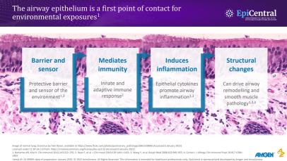
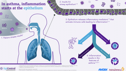
Role of the epithelium in asthma

The airway epithelium has a fundamental role in airway inflammation and asthma.1

The airway epithelium has a fundamental role in airway inflammation in asthma and other respiratory diseases1,2

  • In a healthy state, the airway epithelium is a highly regulated structure providing an efficient physical barrier to environmental exposures.3 It also acts as an immune barrier through controlled recruitment and activation of immune cells; thus, it can be considered an immune-functioning organ4,5​
  • In respiratory disease pathophysiology, the airway epithelium plays a critical role, mediating immunity via ​both the innate and adaptive responses1–4,6,7​
  • The airway epithelium is the first point of contact for environmental triggers, including pathogens, aeroallergens, and irritants1,3,4 ​
  • When inhaled triggers come into contact with the airway epithelium, epithelial cytokines TSLP, IL-33 and IL-25 are released, initiating a cascade of immune responses leading to inflammation and structural changes that contribute to the clinical features of respiratory disease4–6

Increased understanding of the functions of the airway epithelium in healthy and disease states will contribute to better diagnostics and treatment options, to help achieve better patient outcomes2

IL, interleukin; T2, type 2; TSLP, thymic stromal lymphopoietin​
1. Heijink IH, et al. Clin Exp Allergy 2014;44:620–630; 2. Hiemstra PS, et al. Eur Respir J 2015;45:1150–1162; 3. Holgate ST. Immunol Rev 2011;242:205–219; 4. Bartemes KR, Kita H. Clin Immunol 2012;143:222–235; ​5. Roan F, et al. J Clin Invest 2019;129:1441–1451; 6. Gauvreau GM, et al. Expert Opin Ther Targets 2020;24:777–792; 7. Cohen L, et al. Am J Respir Crit Care Med 2007;176:138–145

What is the role of the airway epithelium in its healthy state and in asthma? 

In a healthy state, the airway epithelium is a highly regulated structure consisting of closely bound cells, which provides an efficient physical barrier to environmental exposures from the outside world.3 As well as a physical barrier, the epithelium acts as an immune barrier to the external environment through the controlled recruitment and activation of immune cells.2,4 

Video: Watch Professor Celeste Porsbjerg introduce​ the critical role of the epithelium in severe ​asthma (01:06)

The epithelium also has a fundamental role in asthma pathophysiology,1–3,5,6 as it mediates immunity via both innate and adaptive responses.2 In patients with asthma, exposure of the airway epithelium to environmental triggers results in dysregulation of the epithelium, inducing the release of epithelial-derived cytokines, or alarmins.2,4 In response to cytokine release, there is aberrant infiltration and activation of immune cells leading to chronic inflammation.2,4 In addition, some triggers may alter or damage the epithelium, promoting structural changes that can drive airway remodelling.2,5 Both airway inflammation and remodelling are key characteristics associated with asthma.2,5 

Video: Watch Professor Celeste Porsbjerg ​ discuss how the airway epithelium is the first point ​ of contact for environmental exposures (00:39)

How do inhaled triggers interact with the epithelium?

The epithelium may encounter several inhaled triggers, including pathogens (eg, respiratory viruses or bacteria),7,8 aeroallergens (eg, pollen, house dust mites, animal dander and mould)9 and irritants (eg, cigarette smoke or air pollution, such as diesel particles).10 Triggers among patients with asthma can be diverse, and patients reporting a high burden of triggers can experience more severe asthma exacerbations than those with a low burden.11 

The proximity of the airway epithelium to the external environment means that it needs to respond quickly to stimuli. To enable this quick response, a range of receptors are expressed on the epithelium, including:2,12 

  • Protease-activated receptors (PARs) – these are triggered by stimuli with proteolytic activity, such as fungi and house dust mites
  • Pattern recognition receptors (PRRs) – these recognise danger- and pathogen-associated molecular patterns (DAMP and PAMP, respectively); both are molecules released in response to airway pathogens
    • Toll-like receptors (TLRs) – a type of PRR that interacts with microbial components of airway pathogens

Damage to the epithelium from mechanical injury or irritants, such as smoke, may also cause dysregulation of the barrier functions and downstream inflammation.2

Video: Watch Professor Celeste Porsbjerg explore how asthma triggers interact with the airway epithelium resulting in asthma exacerbations (01:06)

How does the epithelium contribute to asthma pathology and symptoms?  

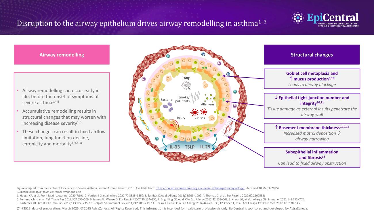
In asthma, as well as marked airway inflammation, there can also be associated structural changes to the airway epithelium (Figure 1), which renders the airways more vulnerable to infection and environmental triggers.2 Both the extent of inflammation and structural changes influence the severity of the disease and asthma symptomatology.3  

Structural changes include goblet cell hyperplasia2,3 and, in more severe disease, a change in mucin expression, primarily increase in the MUC5AC to MUC5B ratio, results in an MUC5AC-rich gel that tethers to epithelial mucous cells and markedly impairs mucociliary transport.13 This increase in submucosal goblet cells and mucus plugging leads to airway blockage.3,13  

There is also a decrease in the number and integrity of tight junctions,1,3 leading to tissue damage as external triggers are able to penetrate the airway wall. 

Increased epithelial thickness2,6 and subepithelial fibrosis6 have also been observed, resulting in airway narrowing and fixed airway obstruction, respectively.  

Finally, there are increased levels of inflammatory cells (including mast cells and eosinophils),14,15 which, in turn, cause heightened inflammation and airway hyperresponsiveness. 

Image
Figure 1: The airway epithelium is altered in asthma

Figure 1: The airway epithelium is altered in asthma.1–3,6,16 Figure adapted from Heijink IH et al. Allergy. 2020;75:1902–1917 (with permission granted under CC license)

The mechanisms contributing to the loss of airway epithelial barrier function need to be elucidated further.14 However, it is well understood that the altered airway epithelium structure allows submucosal infiltration of inhaled triggers that interact with immune cells, causing increased inflammation and associated asthma symptoms.14,16

How does the epithelium mediate airway inflammation? 

When inhaled triggers come into contact with the airway epithelium and specific receptors, epithelial cytokines, referred to as alarmins, are released.5 For example, inhaled microbes can activate TLRs on the epithelial surface, which in turn may cause epithelial cells to produce and release thymic stromal lymphopoietin (TSLP), interleukin (IL)-33 and IL-25.2 In addition, mechanical injury to the airway epithelium may result in release of IL-33.2  

Release of epithelial cytokines initiates a cascade of immune responses that result in inflammation and contribute toward the clinical features of asthma.2,5 Different triggers (eg, allergens, viruses, air pollutants), and subsequent cytokine release, may also result in unique patterns of inflammation (eg, allergic eosinophilic, non-allergic eosinophilic, or non-eosinophilic).5 Inflammation patterns may vary over time and in different situations, within an individual patient; therefore, changes in activated pathways may partly explain the heterogeneous and dynamic nature of asthma.5 

Find out more about the EpiCreator – Professor Celeste Porsbjerg

RELATED RESOURCES

Like this page? We have a host of other resources available in the ‘Scientific and Resource Library’ where you can find out more.

References 

1. Heijink IH, et al. Clin Exp Allergy. 2014;44:620–630. 2. Bartemes KR, Kita H. Clin Immunol. 2012;143:222–235. 3. Holgate ST. Immunol Rev. 2011;242:205–219. 4. Roan F, et al. J Clin Invest. 2019;129:1441–1451. 5. Gauvreau GM, et al. Expert Opin Ther Targets. 2020;24:777–792. 6. Cohen L, et al. Am J Respir Crit Care Med. 2007;176:138–145. 7. Wark PA, Gibson PG. Thorax. 2006;61:909–915. 8. Iikura M, et al. PLoS One. 2015;10:e0123584. 9. Baxi SN, Phipatanakul W. Adolesc Med State Art Rev. 2010;21:57–71. 10. Lambrecht BN, Hammad H. Nat Immunol. 2015;16:45–56. 11. Price D, et al. J Asthma. 2014;51:127–135. 12. Frey. A, et al. Front Immunol. 2020;11:761. 13. Bonser LR, et al. J Clin Invest. 2016;126:2367–2371. 14. Calvén J, et al. Int J Mol Sci. 2020;21:8907. 15. Altman MC, et al. J Clin Invest. 2019;129:4979–4991. 16. Heijink IH, et al. Allergy 2020;75:1902–1917