Complexity of severe asthma

Inflammation in asthma is complex and heterogeneous, which makes it challenging to manage.1–4

Article
 

Overlapping and changing inflammatory pathways in response to an exacerbation, trigger or treatment change highlight the complexity of severe asthma.1–4

Inflammation in severe asthma  

Inflammation in asthma leads to increased symptoms.1,5 Asthma-associated inflammation is complex and heterogeneous,1–4 and numerous cell types, mediators and downstream immune pathways are involved.1,3–6 Multiple inflammatory endotypes have been characterised – allergic and eosinophilic inflammation, to name a few.5 

Professor Christopher Brightling discusses the complexity of severe asthma.

Watch Video


Image
Epicentral Inflammatory pathways
Multiple inflammatory pathways underpin the complexity and heterogeneity of inflammation in severe asthma

Overlapping inflammatory pathways in severe asthma 

Activation of the airway epithelium by exposure to a pollutant or allergen results in the production of epithelial cytokines and leads to a cascade of events that result in airway inflammation and the clinical manifestations of asthma.1,7 This airway inflammation drives changes in asthma pathophysiology and leads to airway hyperresponsiveness and airflow obstruction.1 

The production of epithelial cytokines leads to multiple downstream immune pathways in patients with severe asthma, including:1 

  • Type 2 (T2) inflammation, typically marked by allergic and/or eosinophilic inflammation1 

  • Mechanisms beyond T2 inflammation, characterised by neutrophilic or paucigranulocytic inflammation, and airway hyperresponsiveness, airway remodelling or microbial dysbiosis, which may exist together with T2 inflammation1,8,9 

While many patients have T2 disease,1,2 a sizeable group has mechanisms that are beyond T2 disease.2  

Professor Ian Pavord explains the asthma heterogeneity and the dynamic nature of airway inflammation in asthma.  

Watch Video

Some patients have concurrent overlapping inflammatory pathways.1–4 Often the extent of overlap is unknown; however, it is likely that many patients have a mixture of pathways that drive their disease.1,2 The dominant pathway may change over time owing to different circumstances, such as changes in medications, treatment adherence, exacerbations or exposure to allergens.1–4 In a biomarker study conducted in patients with severe asthma, the phenotype changed in ~50% of patients based on their sputum biomarker clustering after 1 year of follow-up.4  

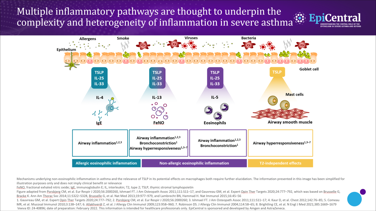
Upstream activation of epithelial cytokines can lead to initiation of multiple downstream inflammatory pathways.10 The wide spectrum and overlap of downstream pathways in severe asthma mean that diagnosis and treatment can be challenging.2 For example, in one US study, around a third of adults with severe asthma have both an allergic and an eosinophilic (defined as blood eosinophils >300 µL/cells) phenotype.2 It would be advantageous to further understand any overlap and common drivers (such as epithelial cytokines) of the downstream endotypes of severe asthma.11,12 Understanding this could help to identify appropriate treatments to address the overlap of downstream pathways and to improve patient care.6,11   

Biomarkers in severe asthma  

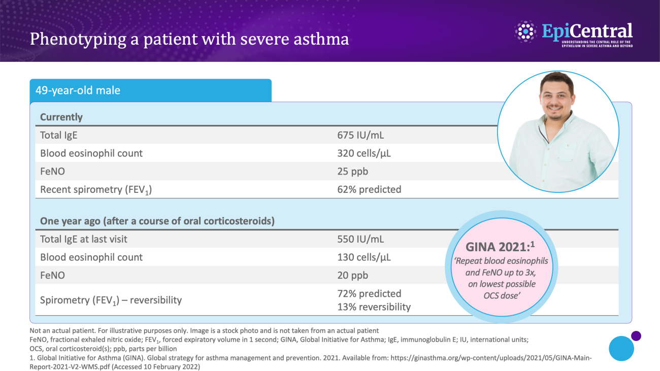
Dynamic, objective and diagnostic biomarkers can help to identify phenotypes and endotypes of severe asthma and help in the selection of an appropriate treatment.13 As such, it is important that clinicians are able to interpret biomarkers effectively to improve patient outcomes.13  

Various biomarkers of T2-mediated inflammation, including specific blood immunoglobulin (Ig) E, blood or sputum eosinophils, and fractional exhaled nitric oxide (FeNO), are available to clinicians1,13,14 and these, among others, can be used in clinical practice for phenotyping of severe asthma.13 

  • Allergen-specific blood IgE levels, measured via a blood or skin prick testing, are higher in patients with allergic asthma compared with healthy individuals and can be used as a surrogate measure for atopy13,15 

  • Blood eosinophils and sputum eosinophils are surrogate markers of T2-inflammation and the T2-inflammatory cytokine, interleukin (IL)-5, which is required for eosinophil activation and survival.14 These are useful biomarkers as patients with higher eosinophil counts are prone to experiencing severe disease and poorer asthma outcomes than patients with lower eosinophil counts16 

  • FeNO is a biomarker of airway epithelial cell exposure to IL-13 and IL-4.10,14 These cytokines upregulate inducible nitric oxide synthase (iNOS) in the airway epithelium, and result in increased nitric oxide production.14 A high FeNO measurement correlates with airway eosinophilia in asthma and indicates increased airway T2 inflammation14 

Image
Case study image example of a typical patient with severe asthma
Biomarker assessment in a 49-year-old man with severe asthma
Image
Case study image example of a typical patient with severe asthma
Phenotyping a patient with severe asthma

It is important to note that biomarker levels may be affected by treatments.15 Biomarkers of T2 inflammation are often suppressed by inhaled corticosteroids and oral corticosteroids; therefore, eosinophils and FeNO assessments are encouraged before commencing a short course or maintenance OCS, or on the lowest possible OCS dose.15  

Some gaps exist in the clinical predictive value of existing biomarkers owing to the challenge of identifying a single predominant endotype of severe asthma.6 Furthermore, there are currently no readily available biomarkers in clinical practice that identify T2-independent asthma.10,17,18 For now, biomarker data need to be interpreted alongside symptoms and lung function, and need to focus on identifying tractable features of asthma, such as airway hyperresponsiveness.19 


Professor Christopher Brightling explains the use of biomarkers in airway inflammation and their predictive value.

Watch Video

Find out more about the EpiCreator – Professor Christopher Brightling

References

1. Busse WW. Allergol Int 2019;68:158–166. 2. Tran TN, et al. Ann Allergy Asthma Immunol 2016;116:37–42. 3. Price D, Canonica GW. World Allergy Organ J 2020;13:100380. 4. Kupczyk M, et al. Allergy 2014;69:1198–1204. 5. Barnes PJ. Pathophysiology of asthma. In: European Respiratory Society Monograph 2003;84–113. 6. Gauvreau GM, et al. Expert Opin Ther Targets 2020;24:777–792. 7. Lambrecht BN, Hammad H. Nat Med 2012;18:684–692. 8. Cao L, et al. Exp Lung Res 2018;44:288–301. 9. Wu J, et al. Cell Biochem Funct 2013;31:496–503. 10. Kuruvilla ME, et al. Clin Rev Allergy Immunol. 2019;56:219–233. 11. Kaur R, Chupp G. J Allergy Clin Immunol. 2019;144:1–12. 12. Agache I, et al. Allergy 2012;67:835–846. 13. Carr TF, Kraft M. Ann Allergy Asthma Immunol 2018;121:414–420. 14. Peters MC, et al. Curr Allergy Asthma Rep 2016;16:71. 15. Global Initiative for Asthma (GINA). Global strategy for asthma management and prevention. 2021. Available from: https://ginasthma.org/wp-content/uploads/2021/05/GINA-Main-Report-2021-V2-WMS.pdf (Accessed 10 February 2022). 16. Kostikas K, et al. Curr Drug Targets 2018;19:1882–1896. 17. Schleich F, et al. Curr Top Med Chem 2016;16:1561–1573. 18. Quoc QL, et al. Exp Mol Med 2021;53:1170–1179. 19. James A, Hedlin G. Curr Treat Options Allergy 2016;3:439–452. 

Read next: Role of the epithelium in asthma

To learn more about how the epithelium mediates airway inflammation, visit the 'Role of the epithelium in asthma' page.

RELATED RESOURCES

Like this page? We have a host of other resources available in the ‘Scientific and Resource Library’ where you can find out more.